VII./2.1.: Main case data of the presented dissection, VII./2.2.: Description of the preparation
 |
VII./2.1.: Main case data of the presented dissection
|
Age of the patient:
|
80 years
|
|
Gender of the patient:
|
Female
|
|
Basic pathology:
|
Pyelonephritis acuta abscedens
|
|
Pathological complication:
|
Abscessus pulmonis sinistri
|
|
Pathological cause of death:
|
Sepsis
|
|
Pathological comorbidity:
|
Meningeoma
|
|
Histopathological diagnosis:
|
Meningeoma meningotheliale
|
VII./2.2.: Description of the preparation
|
 |
The brain weighs 1350 g, on its surface gyri and sulci are intact. On the basal surface, under the left hemisphere, a round, knobby but relatively smooth tumor can be observed, when pushing aside the brain stem. The size of the tumor is 4,2 x 3,5 cm. The tumor is separated from the brain tissue, but almost half of it causes an impression in the substance of the base of the brain, and thus, it reaches the basis of the left ventricule as well. Otherwise, structures and vessels of the base of the brain are usual.
|

Look into the picture!
|
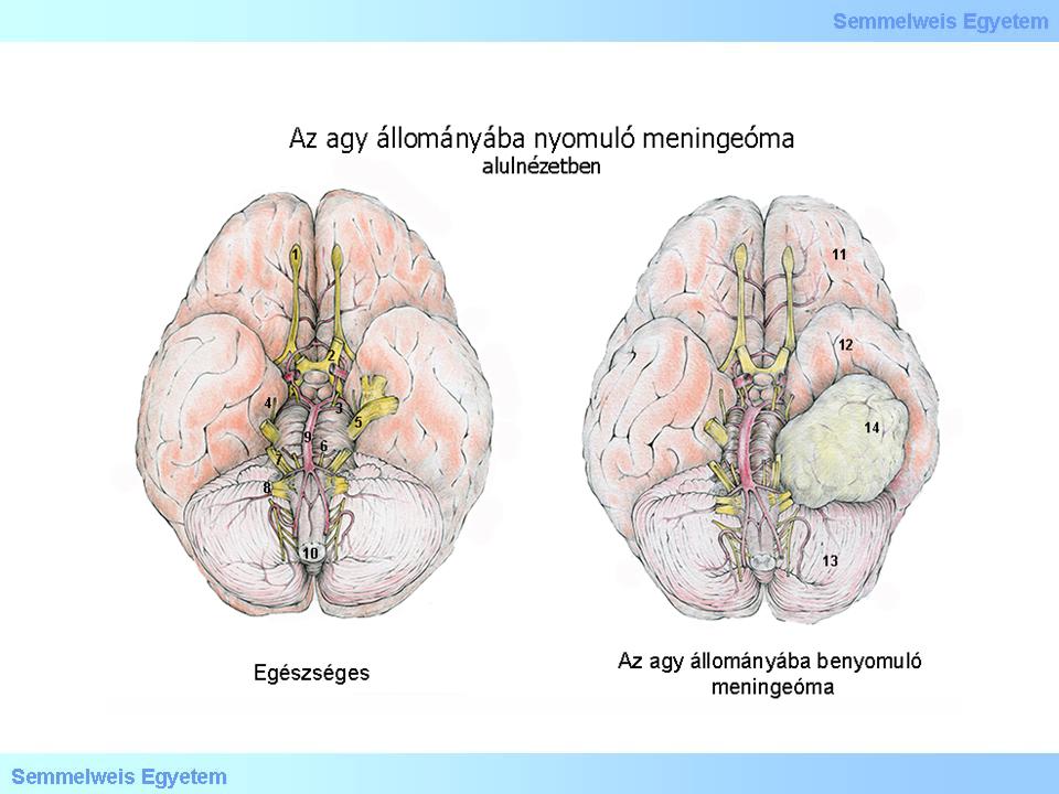
Illustration 1: (1) Nervus olfactorisu et bulbus olfactorius; (2) Nervus opticus et chiasma opticum; (3) Nervus oculomotorius; (4) Nervus trochlearis; (5) Nervus trigeminus; (6) Nervus abducens; (7) Nervus vestibulocochlearis et nervus facialis; (8) Nervus glossopharingeus,nervus vagus, nervus accessories et nervus hypoglossus; (9) A. basilaris; (10) Medulla oblongata; (11) Lobus frontalis; (12) Lobus temporalis; (13) Cerebellum; (14) Meningeoma.
|
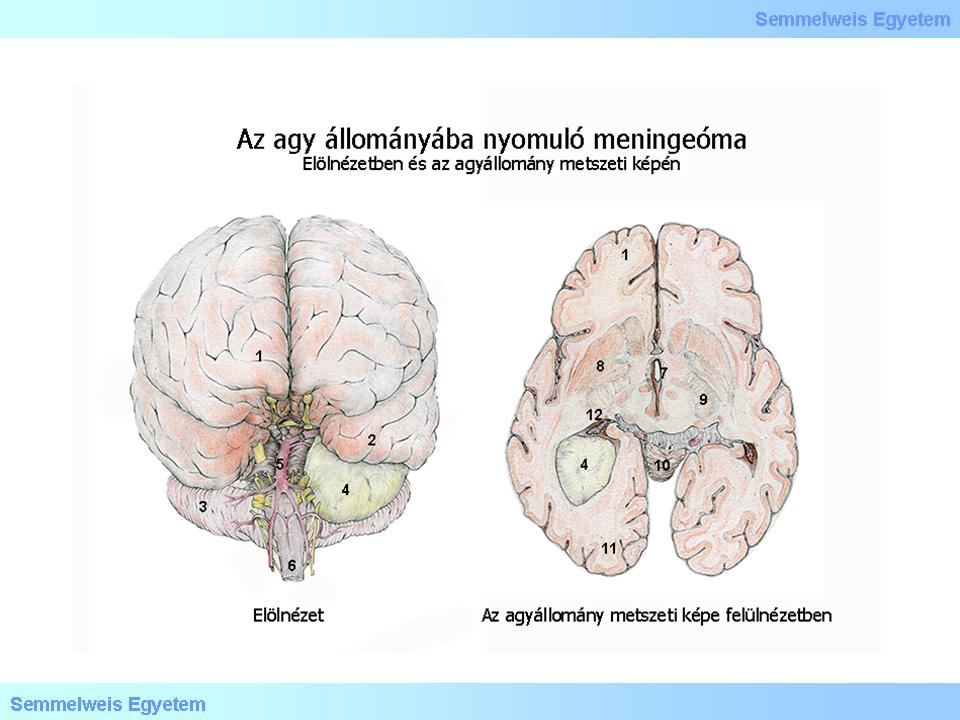
Illustration 2: (1) Polus frontalis; (2) Polus temporalis; (3) Cerebellum; (4) Meningeoma; (5) Arteria basillaris; (6) Medulla oblongata; (7) Ventriculus tertius; (8) Basalis ganglionok; (9) Capsula interna; (10) Vermis; (11) Polus occipitalis; (12) Trigonum ventriculi et plexus choroideus ventriculi lateralis.
|
|
|
Utolsó módosítás: 2014. April 30., Wednesday, 11:08