| |
II./3.3.: Role of CT and MR in the diagnostics of aortic defects
|
 |
The invasive cardiac catheterization and angiography have receded in the diagnostics of the heart defects since the advent and astonishing development of echocardiography; these are currently used only in selected cases in the diagnostics of defects with ambiguous severity. Nevertheless, this technique is eminently suitable for measuring intracardial pressures and valvular gradients, for calculating valvular and regurgitation orifices, regurgitation fractions, as well as for characterizing the heart’s pump function and for measuring the cardiac output. Of course, preoperative coronary angiography has lost nothing of its importance due to the mapping of coronary lesions which require a potential bypass.
Although echocardiography is our number one diagnostic method for examining valvular diseases at the beginning of the 21st century; in a part of patients the exact assessment and accurate quantification of the given lesions may be limited due to the patients’ constitutional-anatomical proprieties, a weak echo window or complex structural cardiac anomalies. In these cases other imaging methods can be used, which are much more expensive, and the cardiac CT examinations are also associated with radiation exposure.
A typical cardiac CT examination includes the recording of at least three series of images, i.e. native, with contrast media, and after contrast media. All of these are done during several cardiac cycles with ECG gating, therefore a significant tachycardia and arrhythmias may lead to artifacts making the evaluation of the valves difficult. In the assessment of aortic defects, the currently established field of application for CT includes the determination of the pathogenic mechanism and severity of aortic stenosis.
|
 |
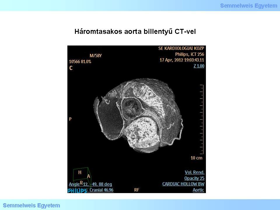
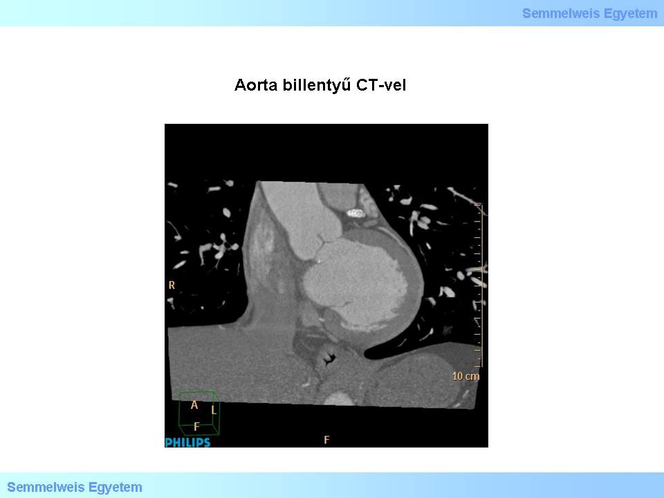
In contrast to echocardiography, the stenotic valvular area can be measured very accurately by planimetry even in patients with a significantly calcified valve and weak left ventricular function. Determination of the valve’s calcium content (calcium score) provides important additional information which predicts the expected rate of disease progression and the increased risk of cardiovascular events. Of course, CT examination is very useful also in the recognition of the malformations of the cusps (Images 6, 7 and 8). Hemodynamic consequences of the stenosis, the left ventricular hypertrophy, changes in its size and shape, as well as the post-stenotic aortectasis can be measured with great accuracy. Before a transcatheter valve implantation, in addition to the cardiac examination also CT angiography is made on the complete thoracic and abdominal vascular beds in order to evaluate the anatomical eligibility.
|

Please, view the images
|

Image 6: Tricuspid aortic valve with CT. Short-axis recording.
|

Image 7: Aortic valve with CT. Long-axis recording.
|

Image 8: Bicuspid aorta with CT. Short-axis recording.
|
In the three-dimensional reconstructed images the assessment of aortic root geometry and vascular angulations is the most accurate with CT. The signs of a heart failure that develops due to the valve’s disease, such as interstitial pulmonary edema, pleural fluid and ascites are clearly visible. CT is also excellent for detecting the dysfunction of implanted artificial valves, and if required in the patient group with a moderate risk it may replace the invasive coronary angiography before surgery. In the differential diagnostics of acute chest pain the „triple rule-out” examination may separate aortic dissection, pulmonary edema and acute coronary syndrome in a single session.
|
 |
As the imaging with CT is not dynamic, the flows cannot be visualized and measured directly. This examination is very accurate for recognizing the etiology of aortic insufficiency; however it cannot be used to determine the extent of regurgitation.
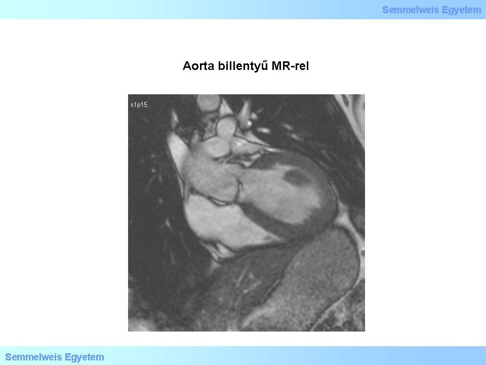
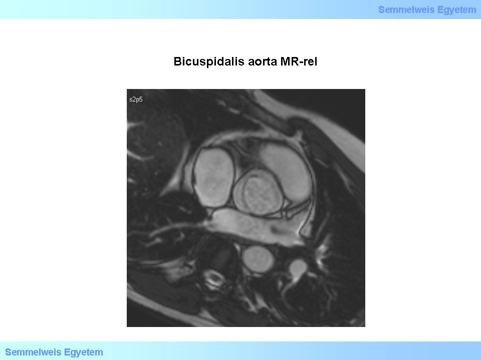
The cardiac MR examination is suitable for the assessment of aortic stenoses even in several ways (Images 9 and 10). The valvular area can be measured by direct planimetry (contouring of the valvular orifice in a cross-sectional plane). Similarly to Doppler echocardiography, MR can also be used for measuring velocities through the stenosis, and a pressure gradient can also be calculated to a sufficient accuracy with the same physical formula; however this is done rarely. The cardiac complications of aortic stenosis, e.g. the increase of the left ventricular myocardial mass and the impairment of systolic function can be measured excellently.
|

Please, view the images
|

Image 9: Aortic valve with MR. Long-axis recording.
|

Image 10: Bicuspid aorta with MR. Short-axis recording.
|
Cardiac MR examination is the imaging method of choice for the follow-up of the aortic dilatation in young patients with an aortic insufficiency, as it is much more accurate than echocardiography, it can depict the entire thoracic aorta and it is associated with neither iodine-containing contrast agent nor ionizing radiation. Allergic reactions to MR contrast media are very rare. MR is also excellently suitable for examining the structure of the thoracic aorta’s vascular wall, e.g. for detecting an edema that developed in the vascular wall due to an inflammation, so that on the base of it, in addition to the artificial valve implantation, reconstruction of the aorta may also be performed during an extended surgery.
|
 |
In patients with aortic insufficiency, in addition to the examination of the valve’s anatomy and the pathology of the great thoracic vessels, MR also provides the most accurate measurement data on the size of cardiac chambers and on the pump function of the ventricles; it is considered the gold standard in this. The signs of regurgitation are depicted well; the measurement or calculation of the regurgitating volume is of approximate accuracy. MR examination of the valvular lesions is rather cost- and time-consuming; it is used only in selected cases.
Both CT and MR play a very important role in the examination of valvular anomalies which occur in the complex cardiac malformations.
|
|