| |
II./2.3.: Meningomyelocele
II./2.3.1.: Meningomyelocele as a form of spina bifida, and other neural tube defects
The neural tube defects (craniospinal dysraphias) may affect different regions of the central nervous system.
|
 |
-
(i) Anencephaly (cranioschisis) is the lack of the brain skull and consequently the forebrain. Anencephaly may occur in itself, but it is commonly associated with cervical, thoracic or total spina bifida. The lack of the brain skull is the primary step in its pathogenesis, and the destruction of the brain tissue is secondary: at first the brain is „free-floating” in the amniotic fluid, later it perishes, and is replaced by vascular connective tissue (area cerebrovasculosa), thus, the typical anencephalic state evolves which is incompatible with life.
-
(ii) If the neural tube defect affects the frontal bone too, the union of the facial buds may be inadequate ("midline facial defect"), which is associated with nasal cleft and face deformity of varying severity due to hypertelorism (hypertelorism: abnormally increased distance between double body parts, especially the eyes. The distance between the eyes is described by the distance between the two pupils. A between-pupil distance of more than 65 mm in adult females and more than 70 mm in adult males can be considered as hypertelorism. A larger distance between the eyes itself is not a disease – to some extent it can even give a kind of appeal to the face [e.g. the case of Liza Minnelli or Jacqueline Kennedy Onassis] – however, it may be a part of several malformation syndromes.)
-
(iii) Spina bifida (split spine, rachischisis) is the openness of the vertebral canal to varying extent. In most of the cases the open state of the vertebral canal and its surroundings results in dorsal protrusions, however, less commonly the protrusion may be abdominal (ventral, or pelvic), or side (lateral, or paravertebral). The protruding bulges of the ventral closing defects are facilitated by sacral defects, while the lateral ones are associated with so-called hemivertebrae. An explicit form of the posterior closing defect is the case when the vertebral meninges protrude by themselves (meningocele) or together with their spinal tissue content (meningomyelocele). If the protruding tissue compasses a cystic cavity which communicates with the central canal (canalis centralis) of the spinal cord, the lesion is called syringomyelocele (myelocystokele).
|
 |
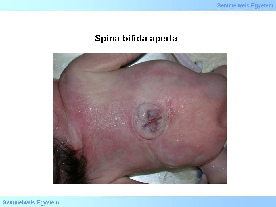
Unfortunately the two latter lesions are commonly associated with explicit and untreatable malfunction of the lower extremities and urinary bladder dysfunctions. Sometimes the protrusion is accompanied by the local increment of mature adipose tissue („lipomeningocele”). Spina bifida disease most commonly affects the lumbosacral section. It is often associated with hydrocephalus, and ventriculomegaly noticed intrauterine may draw attention to it. Partly due to this, and partly due to the paraplegia/paraparesis, incontinentia and risk of urinal tract infections after birth, it has an unfavourable prognosis. In case of spina bifida aperta (open split spine) the vertebral canal is completely open, or only covered by thinned and pathologically structured meninges, the skin surface is breached, and the bare spinal cord or the replacing pathologic nerve tissue can be seen through the usually ovoid aperture (Macro picture 1.) This state includes the risk of central nervous system, namely upward spreading meningoventricular infections.
|

Study and evaluate the pictures!
|

Macro picture 1: Spina bifida aperta. This neural tube defect is more easily recognizable compared to its occult variant. Based on this, severe central nervous system infections may develop. (Source: Semmelweis University 2nd Department of Pathology picture archives, collected by Attila Kovacs and Ildiko Szirtes)
|
The skin is intact above the spina bifida occulta (hidden split spine), maybe only the boned vertebral canal is open; in this case it can only be recognized by careful examination. The latter case may have consequences similar to those of the open spina bifida, but with few symptoms (e.g. limited physical capacity of the spine, reoccurring back aches; in some cases the skin’s circumscribed indentation, hyperpigmentation, angioma, lipoma or insular hypertrichosis above the sacral area, etc.) or with no symptoms at all.
Open spina bifida has variants of different severity and form:
-
a) the meningocele is the protrusion of the dura, filled with CSF, not containing any neural;
-
b) if the meningocele contains brain tissue, that is called meningoencephalocele, or if it contains spinal cord tissue: meningomyelocele;
-
c) if the protruding neural tissue is not covered by its meninges, we call the lesion encephalocele, or myelocele;
-
d) Iniencephaly is a complex disorder of the neural tube defects: cervical vertebrae are disintegrated, thus cervical spina aperta and lordosis develop with the fixation of the head in a retroflected position: the so called „star-gazing position” (this is a hyperextension of the neck, due to the fusion of the posterior fossa and the upper part of the cervical neural tube); the neck is short, through the foramen magnum, brain tissue protrudes toward the medulla oblongata (this rare disturbance also includes tight and deformated chest, lung hypoplasia and circulation disturbances);
-
e) Spina bifida may occasionally co-occur with Arnold-Chiari-malformation, which is characterized by a smaller than usual posterior fossa, with steeper wall (platybasia), tentorium cerebelli in a lower position, elongated pons and medulla oblongata moved toward the foramen magnum, sometimes even broken too, and protrusion of parts of the medulla oblongata and sometimes even the cerebellum, through the foramen magnum, to the spinal tube. The consequent stenosis of the cerebral aqueduct causes obstructive hydrocephalus (this complex aberrant anatomy of the region of the medulla oblongata is in the background of the frequently occurring sudden death of patients with Arnold-Chiari-syndrome).
|
 |
(iv) Neural tube defects may be herited in a multifactorial way, but teratogenic damage can as well, be in the background.Their prevalence rate changes across populations, in western countries it has decreased to cca 1.5‰ in the past decades, due to, most probably, the multivitamins, regularly taken periconceptionally, or the change in nutrition (increased floic acid intake). In these days, fetuses with severe neural tube defect are extremely rarely born, owing to the prenatal diagnostics.
II./2.3.2.: Hydrocephalus and spina bifida
In pathology, hydrocephalus externus and internus are differentiated. In case of fetuses only hydrocephalus internus occurs, with antecedent ventriculomegaly. Over the progressive course of the disease, ventriculomegaly is followed by cortical thinning, then the perimeter of the skull increases, and the fontanelles become gaping. Hydrocephalus is often the consequence of spina bifida, during which obstructive type hydrocephalus develops, frequently with the dysplasia of the Sylvian aquaeduct. Apart from spina bifida, gliosis, postinfective stenosis (toxoplasma- ,and cytomegaloviral infection), haemorrhage, tumor (anaplastic oligodendroglioma, central neurocytoma, medulloblastoma, angioma, teratoma) disturbance in angiogenesis (aneurysm) can cause obstruction, and thus, hydrocephalus.
|
 |
Certain autosomal dominantly and recessively transmitted diseases, and chromosome aberrations may co-occur with hydrocephalus. Hydrocephalus with an X-chromosome connected transmission needs to be emphasized, in which boy fetuses show the stenosis of the Sylvian aquaeduct, and a typically flectated thumb. Hydranencephaly is distinguished from both ventriculomegaly and hydrocephalus. It is characterized by xantochrom pigmented liquor filling out the skull that is otherwise empty. Occlusion of cerebral arteries is considered to be its cause.
|
|