III/4.5: Diagnostics beyond imaging
| |
III/4.5: Diagnostics beyond imaging
|
 |
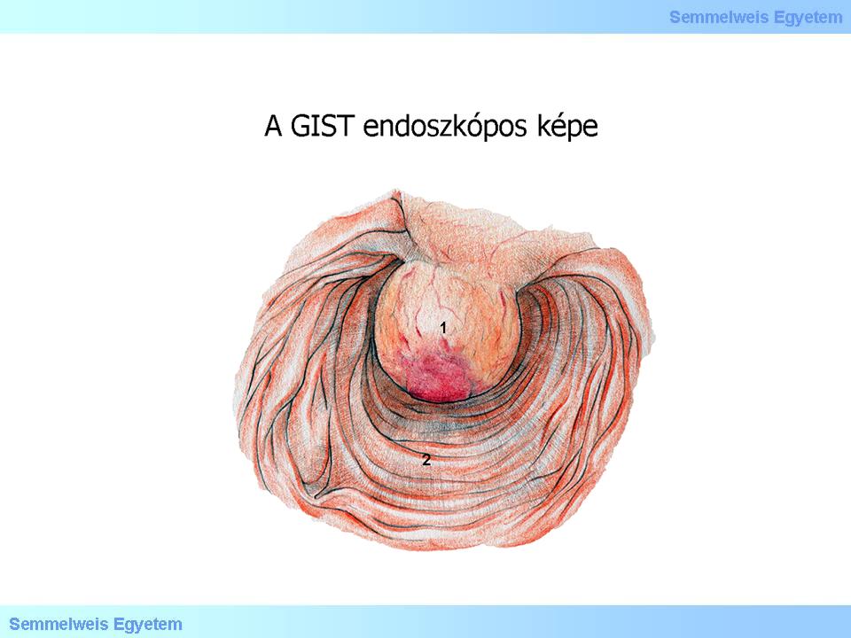
In establishing the diagnosis, the importance of imaging and endoscopic procedures, endoscopic ultrasound examination should be underlined. Nevertheless, it is not easy to recognize GIST during endoscopy: submucosal tumors of small size can hardly be perceived below the usually normal mucosa. A superficial biopsy may also show normal histological conditions. A deeper sample taking or an endoscopic resection is associated with an increased risk of bleeding because of the rich vascular network of the neoplasm. No preoperative biopsy is justified for lesions which can be removed by surgery and are suspicious to be GISTs. Histological examination has a role in confirming a metastasizing process, or prior to the preoperative imatinib therapy of locally advanced GISTs of large size.
When GISTs are localized in the small intestine, the gastroscopy and colonoscopy performed because of a gastrointestinal hemorrhage produce negative results; in these cases conventional enteroscopy, push-enteroscopy, CT enterography, or in case of their unsuccessfulness capsule endoscopy may be taken into consideration: the sensitivity of the latter for GISTs larger than 1 cm is 90 to 100%!

Illustration 1.
|
For determining the exact size of the tumor and its remote metastases further imaging procedures are required such as computer tomography (CT), or magnetic resonance imaging (MRI). In selected cases positron emission tomography (PET) may also be performed; it is also helpful for detecting primary imatinib unresponsiveness.
|
 |
Cells of the tumor express growth factor receptors with tyrosine kinase activity (c-kit), which can be detected by immune histochemical examination (CD-117). CD-117 positivity is one of the most important pathognomonic indicators of GIST; it can be detected in the cytoplasm in 90% of cases.
As blood loss is one of the most characteristic signs of the tumor, anemia of varying severity is often found, particularly in patients with large-size neoplasms.
|
|
Utolsó módosítás: 2014. May 5., Monday, 09:14