| |
II/2.4: Myocarditis
II/2.4.1: General characteristics
|
 |
The clinical picture is characterized by – as opposed to endocarditis – mild initial symptoms or often absolutely no symptoms which is followed by rapid unexpected deterioration in the form of sudden cardiac failure or arrhythmias, or even sudden death. It can affect any age groups, but most frequently young adults. Myocarditis is classified mostly by its pathogenesis: infective; non-infective; idiopathic.
II/2.4.2: Etiology
|
 |
-
- In case of infective myocarditis the most common pathogens causing viral myocarditis are Coxsackie A and B, influenza viruses, Echovirus, EBV, HIV, and CMV. Bacterial myocarditis is most frequently caused by diphtheria bacteria, leptospirae, meningococci, and borrelia (Lyme disease). Protozoa might also be present as pathogens: trypanosoma (Chagas disease) or toxoplasma. Myocardial inflammations associated with rheumatic fever, tuberculosis, or syphilis belong to the so-called specific myocarditis group.
-
- Non-infective myocarditis may occur after physical impacts – like irradiation therapy (ionizing radiation) or electocution. Chemical myocarditis may develop in heavy metal intoxication and as a medication side effect (cytostatics, Sulfonamides, Penicillin). Post-streptococcal myocarditis occurs as part of rheumatic fever. Post-transplant myocarditis is seen during rejection.
-
- Myocarditis of unknown origin (idiopathic) includes giant cell myocarditis, Fiedler’s myocarditis, sarcoidosis associated myocarditis, and eosinophil cell myocarditis which develops on the ground of eosinophil syndromes, allergic processes or viral infections.
II/2.4.3: Morphology
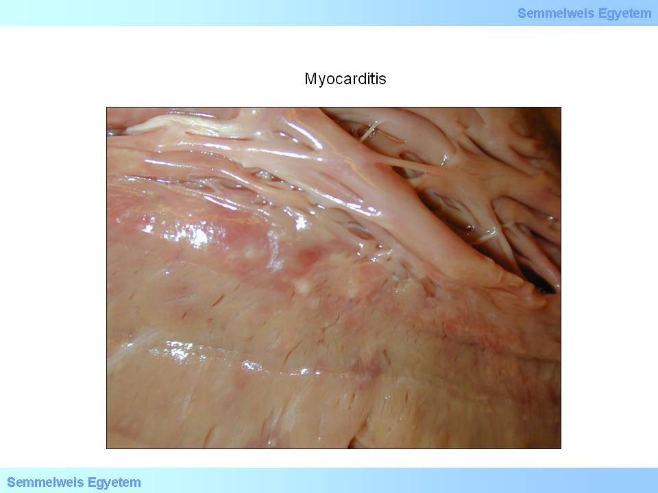
Macroscopically, floppy ventricular dilation is seen, and the myocardial cut surface is dotted with pale, sometimes almost undetectably mottled, small interstitial hemorrhage (macroscopic image No.5). Over a certain extent, atrioventricular valve dilation leads to relative valvular insufficiency thereby exacerbating the sick myocardium’s condition. As for the microscopic image it is important to keep in mind that this is primarily the disease of the interstitium, and myocardial fibre damage – if present at all – is only secondary! Interstitial oedema is always present, and in case of viral myocarditis the infiltration consists of mainly lymphocytes. In other cases – independently from the pathogen – plasma cell, histiocyte and mast cell infiltration is also seen. Later, fibroblast proliferation and consequent interstitial fibrosis develops. Secondary myocardial damage manifests is the form of myocytolysis and microinfarction.
|

Study the image!
|

Mactroscopic image No.5: Myocarditis – macroscopic image is characterized by barely detectable spotting on the cut surface. (From the collection of the 2nd Institute for Pathology, Semmelweis University – collected by Tibor Glasz). b
|
|
|