V./2.1.: Main case data of the presented dissection, IV./2.2.: Description of the dissection
 |
V./2.1.: Main case data of the presented dissection
|
Age of the patient:
|
73 years
|
|
Gender of the patient:
|
Female
|
|
Basic pathology:
|
Arteriosclerosis et hyalinosis arteriarum baseos cerebri
|
|
Pathological complication:
|
Infarctus vetus lobi occipitalis hemispherii sinistri cerebri (pseudoporencephalia partialis)
|
|
Pathological complication:
|
Atrophia cerebri (1A-B. macrographs)
|
|
Pathological cause of death:
|
Thrombosis et occlusio acuta arteriae carotidis internae sinistri
|
|
|
Infarctus acutus lobi frontalis, parietalis et temporalis hemispherii sinistri cerebri
|
|
|
|
|

Look into the pictures!
|
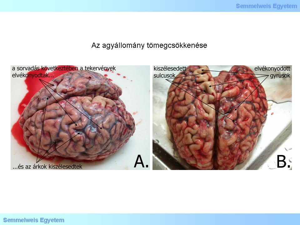
Macrograph 1: Decrease of the brain volume – atrophy – can be caused by various reasons, however, their macroscopic appearance is similar, regardless their orgin: the gyral thinning leads to the flaring of the sulci. (From the image-archive of the Semmelweis University, 2nd Department of Pathology–collection of Attila Kovács)
|
IV./2.2.: Description of the dissection
The brain weighs 1360 g, the structure of the gyri and sulci is usual, at the surface of the occipital pole and the cerebellum, in the subarachnoidal space, there is a blackish red, partly clotted haematoma that covers as a thin layer the surface of the affected areas, and can be observed in the cut-surface of the fourth ventricule too.
|
|
Zuletzt geändert: Wednesday, 30. April 2014, 10:54