|
II./1.3.: The structure of the semilunar (crescent or half-moon shaped) valves
II./1.3.1.: General characteristics of the semilunar valves
The semilunar valves are located in the arterial orifices of the ventricles. It’s important to note that the in- and outflow paths of the right ventricle (the tricuspid and pulmonary trunk orifices and valves) are separated from each other by the supraventricular crest (a myocardium ridge) but in the left side of the heart there is no such muscular structure, the blood inflow and outflow are separated only by the anterior cusp of the mitral valve. At the outflow orifice of the right ventricle the pulmonary trunk valve, at the left ventricle outflow the aortic valve can be found. Both arterial orifices have a valve composed of 3, half-moon-shaped leaflets („pockets” – often called „cusps”) (Fig. 2A-2B. and 4.).
|
 |
Structurally, a denser, stiffer, mainly collagen fiber containing pars tensa and a looser, elastic fiber rich pars flaccida can be distinguished at each semilunar leaflet. The pars tensa has a shape of a one-third sphere; it is like an upwardly opened pocket. Above each pockets there are –at the closure line- 2-2, lunulae (crescent-shaped, looser parts, pars flaccida). Since a round cross sectional area of the vascular lumen cannot be closed completely by 3 pockets like these, the looser, mobile lunulae meet and overlap with each other during valve closure and usually bend towards one side. Between 2 lunulae of a leaflet a nodule is located (pinhead sized, denser connective tissue knots) – providing perfect closure of the valve (fully closing even the geometric centre of the lumen).
Their function: during ventricular contraction (systole), the valve is open, but the leaflets do not reach the wall of the arteries (pulmonary trunk, aorta) since the turbulent blood flow keeps them away from the wall. This fact has a particular importance at the aortic valve since the coronary arteries originate from the aortic sinuses (the dilated portions of the ascending aorta immediately above the semilunar valves) thus allowing the coronary perfusion during systole also. At the ventricular diastole the blood moves backwards in the ascending aorta and pulmonary trunk and thus fulfills the pockets of the semilunar valves, spreading them out, resulting closure of the valve (this situation is even better for the coronary perfusion). The perfect closure is made by the meeting of the adjacent lunulae and the central occlusion of the 3 nodules.
|
 |
The development of the semilunar valves: the left and right semilunar valve leaflets of both vessels arise from common (ie. right and left) endocardium cushions derived from the aorticopulmonary septum; this proliferating endocardium divides the truncus arteriosus into aorta and pulmonary trunk. The anterior semilunar valve leaflet of the pulmonary trunk and the posterior aortic valve leaflet are created by a secondary intimal proliferation (anterior and posterior ones, respectively). The intimal proliferations are initially compact tissues but later cavities are formed in the developing valve tissue. If the tissues of the 3 valve leaflets remain attached to each other in certain areas, congenital valve stenosis (aortic stenosis, pulmonary stenosis) develops, resulting sometimes only a very narrow (pin hole) free lumen.
II./1.3.2.: Pulmonary trunk valve
The pulmonary trunk valve contains 3 semilunar valve leaflets, the anterior, the right and the left ones.
II./1.3.3.: Aortic valve
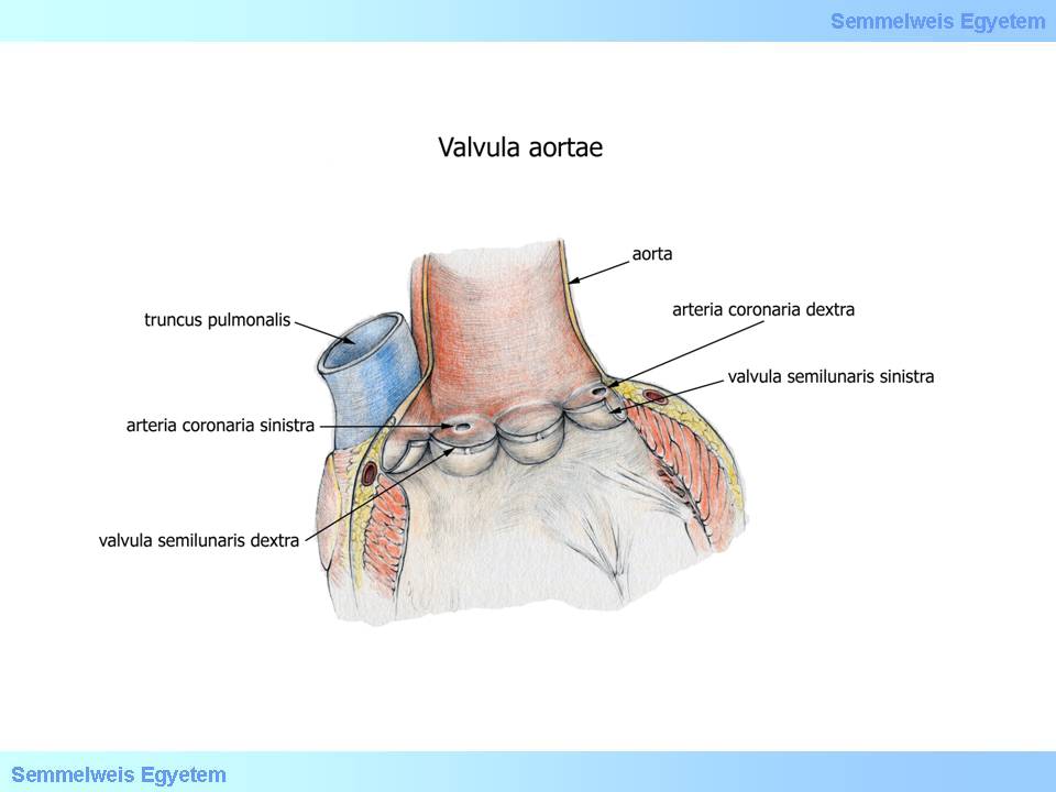
The aortic orifice exhibits the aortic valve formed by the posterior, right and left semilunar leaflets (3 „cusps”, ’pockets’) (Fig. 4.). The aortic wall opposite to the individual leaflets bulges outward (aortic sinus, sinus of Valsalva; collectively resulting the onion-like bulbous dilatation of the ascending aorta: bulb of the aorta).
|

Have a look at the figure and think it over!
|

Figure 4.: The aortic valve and the mitral valve – exposed by opening the left ventricle and the aorta
|
The right and left coronary arteries initiate from the ascending aorta -from the right and left aortic sinuses, at the level of the upper edge of the corresponding valve leaflet. There is no coronary artery arising from the aortic sinus above the posterior valve leaflet (thus called the ’non-coronary sinus’). The coronary arteries practically originate just below the sinotubular junction (border between the wider aortic bulb containing the aortic sinuses and the beginning of the tubular, narrower ascending aorta).
|
|