| |
VII/1.2: General structure of the base of the brain
VII/1.2.1: General remarks
|
 |
It is the most subservient to study the base of the brain on a removed brain conservated in formaldehyde, after removing the remaining soft meninges, namely the pia mater and arachnoid mater. The anterior part of the base of the brain is constituted by the basal surface of the frontal lobe. Behind the frontal lobe, appears the basis of the temporal lobe. This passes into the basal surface of the occipital lobe with a laterally convex hump, continuosly, without a sharp border. The two frontal and temporal lobes frame the interbrain (diencephalon) from the front, sideways and from above, while the two temporal and occipital lobes frame the brainstem and cerebellum sideways and from above.
|
 |

2. fotó: Az agy felszíne az arachniodea eltávolítása után felülről – Molnár Attila és Balogh Attila
|

3. fotó: Az agy felszíne az arachniodea eltávolítása után elölről – Molnár Attila és Balogh Attila
|
VII/1.2.2: Frontal lobe
|
 |
The basis of the frontal lobe is located in the front, in the anterior cranial fossa, and its clinical name is frontobasis. The most anterior point is called frontal pole. Of the gyri we can see the straight gyrus along the longitudinal cerebral fissure in the midline, and laterally from the above are the orbit(ofront)al gyri. The straight gyrus is separated from these by the olfactory sulcus laterally. On brains that were conserved by formaldehyde in situ and taken out later, orbital gyri often show a very concave impression which is caused by the intracranial bulge of the orbital roof. Similarly, orbital gyri also cause characteristic impressions on the orbital roof, and those are called gyral impressions.
|
|

4. fotó: Az agy frontalis basisa – Molnár Attila és Balogh Attila
|
VII/1.2.3: Temporal lobe
The middle cranial fossa is located lower than the anterior, and is filled by the temporal lobe. The most anterior point, reaching the lesser wing of the sphenoid bone is called temporal pole. At the lateral end of the temporal lobe the inferior temporal gyrus turns onto the basis, and further back it is called lateral occipitotemporal gyrus. The two are partly separated by a varying groove which is often missing.
|
|
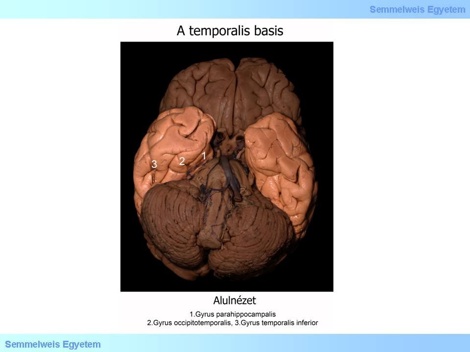
5. fotó: A temporalis basis – Molnár Attila és Balogh Attila
|
More medially, the medial occipitotemporal gyrus is seen, and – similarly to the former gyrus – it reaches the basis of the occipital lobe without a sharp border. The two convolutions are separated by the occipitotemporal sulcus. Medially from the medial occipitotemporal gyrus is a longitudinal convolution called parahippocampal gyrus. They are separated by the collateral sulcus. This also has a varying appearance and is often intermitted by additional convolution parts, mainly on the frontal section. This groove creates a swelling in the lateral cerebral ventricle’s lower (temporal) horn and it is called collateral eminence. The parahippocampal gyrus reaches from the basal to the medial surface of the lobe (this is often called ’mesial’ surface in practice), and its anterior part which has a hook shaped curve is called the uncus of the parahippocamppal gyrus.
VII/1.2.4: Occipital lobe
As we saw before, the occipital lobe doesn’t separate sharply from the temporal lobe, and the temporal gyri continue here with no definitive border. Only an arbitrary border can be made by connecting the parietooccipital sulcus on the medial surface and preoccipital notch on the lower part of the convexity. The small lingual gyrus is seen medially from the posterior end of the occipitotemporal gyrus. In front of, and medially from the lingual gyrus, the posterior part of the parahippocampal gyrus and the collateral sulcus extends a little bit and therefore they form the collateral trigone in the lateral ventricle’s occipital horn. The most posterior point of the occipital lobe is called the occipital pole.
VII/1.2.5: Brainstem
All three parts of the brainstem can be seen from the base: the midbrain (mesencephalon) is on the top, the pons is below it, and medulla oblongata is located caudally. These are ’embraced’ dorsolaterally by the cerebellar hemispheres; therefore the dorsal surface of the brainstem practically remains hidden.
|
|

6. fotó: Az agytörzs – Molnár Attila és Balogh Attila
|
VII/1.2.5.1: Midbrain (mesencephalon)
In the midline of the midbrain is the posterior perforated substance which constitutes the bottom of the interpeduncular fossa. This is bilaterally bordered by the cerebral crura which constitute the basis of the mesencephalon.
VII/1.2.5.2: Pons
More caudally, the pons (pons Varolii) is separated from the midbrain by the pontomesencephalic sulcus. After removing the meninges, the superficial bands of the transverse fibers of the pons and the shallow but wide basilary sulcus on the basilary part of the pons become well visible even with naked eye. The middle cerebellar peduncles (brachia pontis) connect the two sides of the basis pontis with the cerebellum. The cerebellar flocculus is found between the inferior part of the peduncles, the pons and the cerebellum. Below and medially next to the flocculus is a part of the choroid plexus of the fourth cerebral ventricle called Bochdalek’s flower basket.
VII/1.2.5.3: Medulla oblongata
Caudally from the pons and separated by the bulbopontine sulcus, appears the medulla oblongata. On the ventral surface there are two rostrocaudal eminences, the pyramids with the ventral (or anterior) median fissure between them. Lateral to the pyramid is the preolivary sulcus; more laterally is the olivary body and the postolivary sulcus. Behind the latter, another strong band runs in an upward, backward and lateral direction at the posterolateral part of the medulla, and it is called inferior cerebellar peduncle or restiform body. The border between the spinal cord and the medulla oblongata is not sharp. The pyramid continues in the anterior funiculus of the spinal cord, while the inferior cerebellar peduncle continues downward in the lateral funiculus.
VII/1.2.5.4: Interbrain (diencephalon)
The diencephalon can be observed only in a narrow area between the frontal and temporal lobes and the caudally neighbouring mesencephalon. Here we can see a few structures of the hypothalamus, such as the mamillary bodies that border the interpeduncular fossa from the front; the median eminence and tuber cinereum with the pituitary stalk in front of them; and the optic chiasm that continues in the optic nerves rostrally from the above. The optic tracts that start from the chiasm in a posterolateral direction, frame the above diencephalon parts from lateral. They end more dorsally at the lateral geniculate body (CGL) of the methatalamus, but these endings can only be seen with the mutilation of the temporal lobe (parahippocampal gyrus).
|
|