| |
IV./1.3.: Pancreas
IV./1.3.1.: Topography, parts
|
 |
Inseparable from the curvature of duodenum, this gland of 15-18 cm length and 70-90 g weight is adherent to the dorsal abdominal wall. Flattened in a dorsoventral direction, the two surfaces converge in a wedge below. The head (caput pancreatis), which is wider than the other parts, fills the concavity of duodenum. This means that the concave side of duodenum is accomodated, at times nearly enclosed, by the groove of pancreas, tightly anchored by connective tissue. The lower side of the head curves back on itself like a leftward directed hook, following the inferior part of duodenum. This is the uncinate process, separated from the body of pancreas by the pancreatic notch (incisura pancreatis).
Ascending slightly to the left behind the pylorus, and having crossed in front of the inferior vena cava and the aorta, the head continues in the body (corpus pancreatis). The broken line of transition between the head and the body forms a ventrally and superiorly directed elevation termed tuber omentale, where the pancreas is directly related to the lesser omentum. Further ascending slightly to the left, the body gets narrower and continues without a sharp boder in the tail (cauda pancreatis). The latter lies in the left hypochondrium and may, in some cases, reach the hilum of the spleen, with an obtuse ending.
IV./1.3.2.: Peritoneal relations
Given its characteristic retroperitoneal position, only the anterior surface of the pancreas is covered by peritoneum. The omental bursa is situated ventrally to the pancreas, thus the posterior wall the of the bursa corresponds to the peritoneal covering of pancreas (9A-Drawing 1). The transverse mesocolon passes along the anterior surface of the head and the body, with a slight right-to-left elevation. Peritoneal covering of the pancreas is absent under the line of attachment. Since the tail of the pancreas may reach the spleen (a fully intraperitoneal organ), this part of the pancreas may also be enwrapped by peritoneum, i.e. it may become intraperitoneal, situated, in such cases, within the phrenicosplenic ligament.
IV./1.3.3.: Syntopy
In correlation to the spine, the head of pancreas lies at the level of lumbar vertebrae 1-2, on the right side of the vertebral column. The tuber omentale is situated at the border between the thoracic vertebra 12 and the lumbar vertebra 1, in the midline. The body of pancreas passes in front of the lumbar vertebra 1, while its tail lies at the lower border of thoracic vertebra 12, overshooting the level of the left costal arch by 2-3 finger's breadth.
Syntopic relations to other organs:
|
 |
-
- The posterior surface of pancreas is directly adherent to the dorsal abdominal wall;
-
- The head is related to the right crus of the diaphragm and the psoas major muscle;
-
- The body is related to the inferior vena cava and the aorta in front of the spine, as well as to left crus of the diaphragm;
-
- The tail is related, through the renal capsules, to the anterior surface of the left kidney;
-
- Pending sufficient length, the tail of pancreas may reach the hilum of spleen;
-
- Apart from the lower region of the head, the pancreas is related to the dorsal surface of stomach through the omental bursa;
-
- Here, at the tuber omentale, the pancreas is related to the lesser omentum and, thereby, also to the inferior (visceral) surface of the liver;
-
- The head is attached to the concavity of duodenum;
-
- The lower, inframesocolic surface of pancreas is related to the small intestines.
|

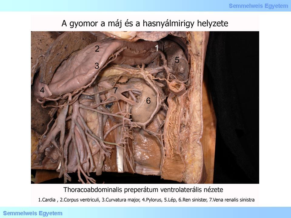
Look at the photo!
|

Photo 3.
|
Topographic relation of the pancreas to the major vessels is of utmost significance. The celiac trunc branches off from the abdominal aorta above the body of pancreas. Its leftward directed branch, the splenic artery passes along the superior border of the body and the tail of pancreas to the spleen. The other branch called common hepatic artery has no contact with the pancreas but its secondary branch, the gastroduodenal artery and its terminal branch, the superior pancreaticoduodenal artery are already deeply embedded into the head of pancreas. The next main visceral aortic branch, the superior mesenteric artery, branches off behind the pancreas. First, this vessel passes in this position, and then, having emerged from the pancreatic notch (incisura pancreatis) between the head and the body, it courses further downward and forward, crossing in front of the horizontal (third) part of duodenum. The latter organ is caught in a fork formed by the superior mesenteric artery together with the aorta. Prior to its crossing with the duodenum, the superior mesenteric artery gives off the inferior pancreaticoduodenal artery, which turns to the right and anastomoses with the superior pancreaticoduodenal artery at the pancreatic head.
This anastomosis is frequently present on both the anterior and posterior sides of the head, rather like a ring.
Typically, the vein accompanying the superior mesenteric artery is tightly apposed to its right side. Having passed through the pancreatic notch, the superior mesenteric vein joins the splenic vein behind the tuber omentale and in front of the inferior vena cava to form the portal vein. The splenic vein passes from the hilum of spleen all the way behind the pancreas, running counter to the splenic artery, which follows the upper edge of the body and tail of pancreas, as far as the spleen. Part of the portal system, the inferior mesenteric vein joins, in most cases, the splenic vein from below, at the transition between the body and tail of pancreas.
IV./1.3.4.: Vascular supply
Vascular supply of the pancreas is derived from two different arterial systems. The branches (rami pancreatici) for the body and tail are supplied by the splenic artery, which belongs to the system of celiac trunc. Branch of the gastroduodenal artery, the superior pancreaticoduodenal artery forms and anastomosis with a direct branch of the superior mesenteric artery, the inferior pancreaticoduodenal artery, to supply the head.
The veins of pancreas drain to the portal vein.
IV./1.3.5.: Excretory ducts
The main excretory duct, the major pancreatic duct (of Wirsung) passes deep near the dorsal surface all the way from tail to head. Here it turns at an obtuse angle downward and pierces the wall of duodenum together with the common bile duct. Then, the duct opens either jointly with the bile duct, or immediately inferior to its orifice, on the major duodenal papilla of the second (descending) part of duodenum. Another smaller duct, arising from the lower part of the head and the uncinate process, the accessory pancreatic duct (of Santorini) crosses the main duct and opens above its orifice on the minor duodenal papilla into the duodenum.
IV./1.3.6.: Lymphatic drainage
Lymph from the pancreas is returned to regional nodes belonging either to the duodenum or to the spleen, all being associated with the celiac lymph nodes.
IV./1.3.7.: Nervous supply
Parasympathetic innervation of the pancreas comes from the branches of vagus nerve, whereas sympathetic innervation is supplied by the celiac plexus, arising from the celiac ganglion.
|
|