I./1.1.: Coronary arteries - in general
| |
I./1.1.: Coronary arteries - in general
Coronary arteries (right and left coronary arteries) deliver blood to the heart musculature (working myocardium and modified conducting cardiomyocytes). The presence of coronary artery disease (CAD) may result in serious consequences by reducing the supply of oxygen and nutrients to the myocardium, which may lead to a myocardial infarction (MI).
|
 |
Coronary arteries originate normally from ostia localized in the left and right aortic valvular sinuses, respectively. The region of the aortic valvular complex displays three sinuses which are dilatations of the wall of aortic root above the attached base of each of the semilunar cusps of the aortic valve. Several alternative names were/are used in clinical practice to refer to the aortic sinus: the sinus of Valsalva, the sinus of Morgagni, the sinus of Otto, or Petit's sinus. Anatomically the three sinuses are different - the left and right sinuses give rise to the coronary arteries, while no blood vessel originates from the third aortic sinus, and hence it can be nominated as the non-coronary aortic sinus.
The coronary arterial ostia are localized usually immediately below or at the sinutubular ridge (supravalvular ridge) within the aortic sinus but above the cuspal margins. The sinutubular ridge is a slight circumferential thickening at the sinutubular junction representing the distal boundary of the aortic root. The left coronary ostium is frequently found in a central position between the commissural attachments of aortic cusps; consequentially, the origin of the right one is often shifted to the right and the left ostium is rarely shifted to the left. In quite a high percentage of hearts, accessory ostia (1 – 4) occur in the right aortic sinus. Rarely, the left and right coronaries originate from the same aortic sinus. A single coronary artery may arise either from the left or right aortic sinus.
The initial segment of coronary arteries, through the wall of the aortic root is funnel-shaped or slit-like. Generally, the size of the left ostium is larger than that of the right.
|

Study the images, and analyze what you see!
|

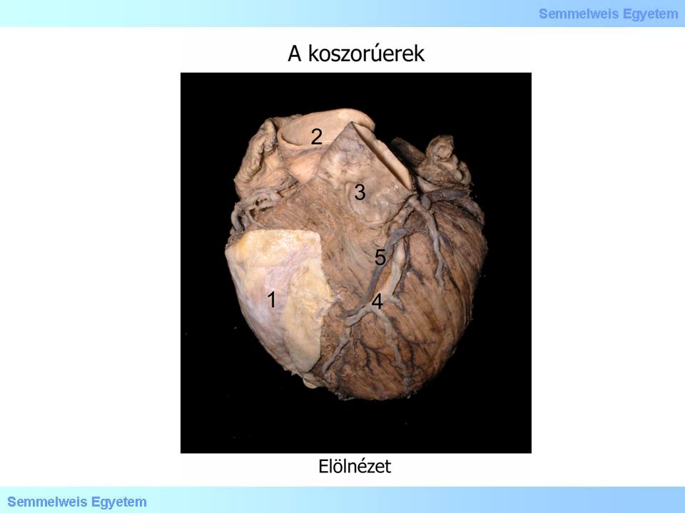
Photo 1.: A koszorúerek előlnézetben – Molnár Attila és Balogh Attila
(1) Pericardium; (2) Aorta; (3) Truncus pulmonalis; (4) Arteria interventricularis anterior ( LAD ); (5) Vena interventricularis anterior
|
The main stems of coronary arteries and their larger branches have an epimural course, i.e. beneath the epicardium, in the subepicardial areolar connective tissue. However, the major branches in the interventricular grooves are often hidden by smaller or larger overlapping myocardial strands (myocardial bridges).
|

Study the photo!
|

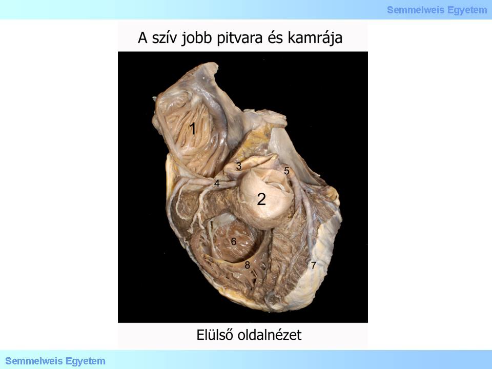
Photo 2.: A szív jobb pitvara és kamrája - Molnár Attila és Balogh Attila
(1) Atrium dextrum ( auricula ); (2) Truncus pulmonalis; (3) Aorta; (4) Arteria Coronaria dextra; (5) Arteria coronaria sinistra; (6) Ventriculus dexter; (7) Pericardium; (8) Trabecula septomarginalis
|
|
|
Last modified: Wednesday, 30 April 2014, 8:41 AM