| |
I./1.2.: Lateral ventricles
I./1.2.1.: General characteristics
|
 |
The two lateral ventricles are the cavities of the forebrain (telencephalon); they follow the brain’s curved outer form with their typical rostrocaudal curvature (Drawing 14A-1.). Each lateral ventricle communicates through a foramen interventriculare Monroei with the third ventricle located in between.
Lateral ventricles consist of four main parts, which correspond to each cerebral lobe’s cavity. Thus the anterior horn (cornu anterius seu frontale) belongs to the frontal lobe, the central part (pars centralis seu cella media) to the parietal lobe, the inferior horn (cornu inferius seu temporale) to the temporal lobe and the posterior horn (cornu posterius seu occipitale) to the occipital lobe. No sharp borderline can be drawn between each part. However, the border between the cella media and the cornu anterius is commonly given as the frontal plane drawn through the foramina interventricularia. The separation of the other ventricle parts is even less possible as they meet in a common, dilated section.
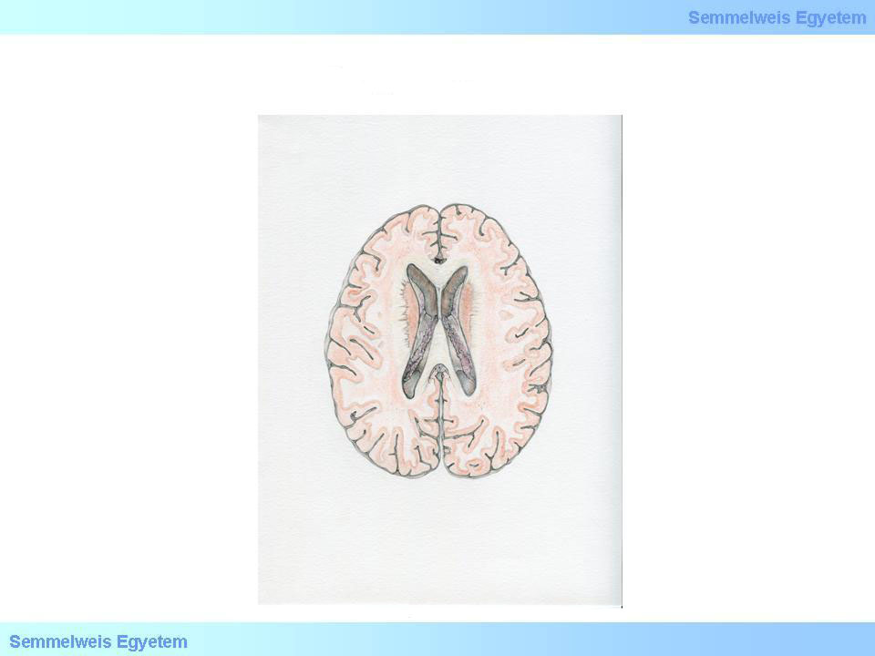
Drawing 1: Horizontal section of the forebrain to illustrate the lateral ventricles
|
I./1.2.2.: Anterior horn (cornu anterius seu frontale)
Its anterior wall is formed by the descending part of the corpus callosum, the genu corporis callosi. Medially it is bordered by a thin membrane, the septum pellucidum, while the above border is again the corpus callosum and its radiating fibers. Its side-bottom wall is formed by the head of the caudate nucleus (caput nuclei caudati). This bulges into the cavity of the ventricle horn to varying degree. In young persons it is quite close to the septum pellucidum, while in elder age a larger gap may appear between them, due to atrophy. The medial wall closely touches the medial wall of the opposite cornu anterius, thus the two septum pellucidum form a membrane duplication. It contains a small cavity, the cavum septi pellucidi.
I./1.2.3/A.: Central part (pars centralis, seu cella media)
The upper wall is again made of the radiation of the corpus callosum. The lateral wall is the body of the caudate nucleus, the corpus nuclei caudati. Structures of the bottom wall from here in lateromedial order are the stria terminalis (containing the v. thalamostriata), the plexus choroideus ventriculi laterales, the dorsal surface of the thalamus with the lamina affixa thalami and the corpus fornicis. The fornix, rising flatly from the former, also constitutes the medial wall.
I./1.2.3/B.: Posterior horn (cornu posterius seu occipitale)
It is the backward-extending, hook-like continuation of the central part. All of its walls are essentially formed by the corpus callosum and its radiation. The lateral wall is also called as tapetum. On its medial wall a prominence called calcar avis appears that corresponds to the medial cerebral surface’s sulcus calcarinus. A thinner, parallel prominence, the bulbus cornus occipitalis can often be found above this. On the bottom of the horn there is an area widening in triangle-shape, called the trigonum collaterale. In a frontal section the sulcus collateralis can be seen on the basis in the opposite side.
I./1.2.4.: Inferior horn (cornu inferius seu temporale)
Starting from the point where the above two ventricle parts meet, it stretches downward, forward and slightly to lateral direction. Its front end approaches the polus temporalis. Embedded between these two, the corpus amygdaloideum can be found in the temporal white matter. The lateral, upper and lower walls of the ventricle part are formed by white matter, while the medial wall is of more complicated construction. In forward direction the hippocampus bulges increasingly into the ventricular cavity. The shallow crosswise grooves on its surface are called sulci interdigitales, and the in-between protrusions are the digitationes hippocampi. In some cases the front part of the hippocampus is not fully separated from the ventricle wall above it, but it is connected by tiny cerebral tissue bridges. We often see that the front end of the hippocampus quasi merges into the temporal lobe’s parenchyma and no free ventricle part remains in front of it. Backwards in top of the hippocampus the covering brain membrane gradually rises in a crest-like shape, forming the fimbria hippocampi. Above it, medially and embedded into the wall the cauda nuclei caudati can be found.
|
|