|
II./2.3.: Myoma or myomatosis uteri
II./2.3.1.: Mesenchymal uterine tumours
Benign mesenchymal tumours of the uterus are rare besides the leiomyoma of smooth muscle; only the benign adenomatoid tumours of mesothelial origin are worth mentioning.
II./2.3.2.: Uterine leiomyoma
|
 |
Myometrial leiomyomata are extremely frequent. They occur in around 25% of women older than 35 years. The tumours are often multinodular. In case of a great number of tumours the condition is called leiomyomatosis. Leiomyomata of the female genital tract are usually tumours of the uterine corpus, sometimes occurring in the cervical or in the tubal muscle wall, or even in the ovarial stroma. Since oestrogen stimulation plays a role in their development, leiomyomata occur typically in the sexually mature age. After the menopause leiomyomata usually settle or retract. Their hormone sensitivity can be used medically: after GnRH treatment their size can reduce, sometimes extended necrosis can develop within them. The leiomyomata can be located directly below the uterine mucous membrane (submucous leiomyomata), deep in the muscular wall of the uterus (intramural leiomyomata), or close to the covering serous membrane (subserous leiomyomata). Subserous tumours can sometimes grow onto the broad ligament of the uterus (ligamentum latum uteri) or may become pedunculated into the abdominal cavity, finally adhering to the omentum or to the peritoneum. If leiomyomata detach from their original connections and get their blood supply from the new location, the phenomenon is called a parasitic leiomyoma. The size of leiomyomata can vary from a few millimetres, grams to more kilograms. Corporal leiomyomata are typically round, their edges are surrounded by a pseudocapsule consisting of squashed uterine muscle (1st macrophoto).
|

Please take a look at the pictures!
|
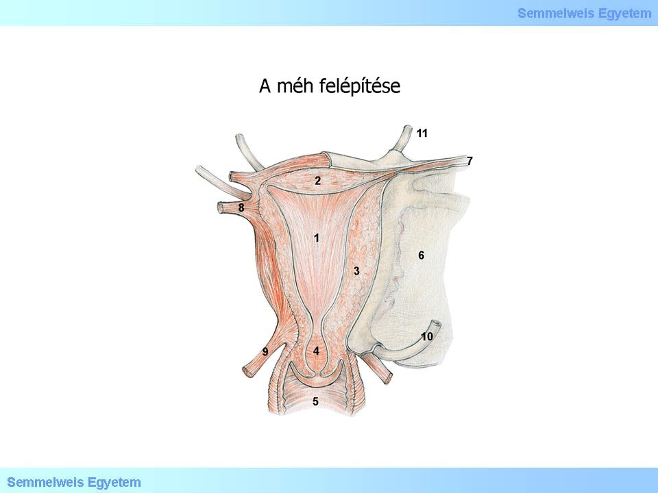
1st illustration : Uterus myoma (Kiss Balázs): (1) Peduncularis subserosus myoma; (2) Subserosus myoma; (3) Intramuralis myoma; (4) Intracavitalis myoma; (5) Subserosus myoma; (6) Prolapsus myoma
|
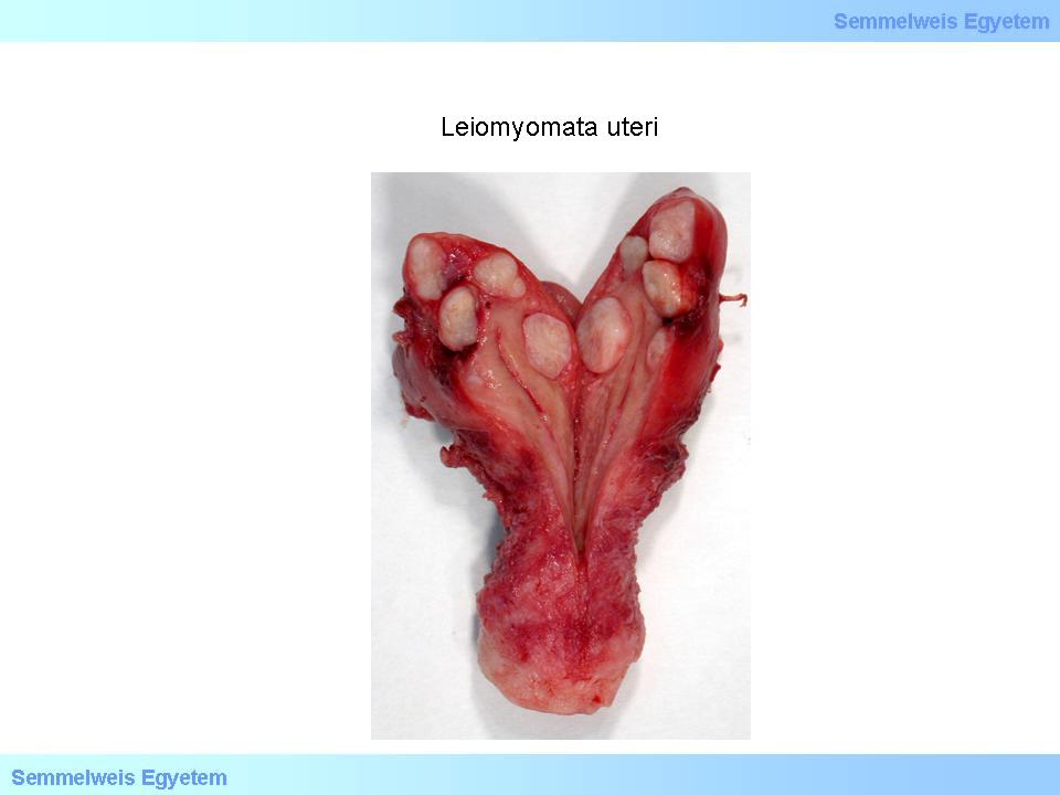
1st Macrophoto: Leiomyomata uteri. Multiple benign intramural or subserous smooth muscle nodules with a characteristic cut surface of whirl-like fibres (From the photo archive of the Semmelweis University 2nd Institute of Pathology – collected by Attila Kovács and Ildikó Szirtes).
|
Their structure consists most often of whirly, white bundles on the cut surface. The tumour tissue is has a solid feel. Bigger leiomyomata can contain bleedings, thinning and myxoid degradations. Histologically smooth muscle cells forming interwovened bundles are present. Smooth muscle cells have a distinctively elongated nucleus with rounded ends, which resemble a cigar (1st micropicture).
Macroscopically the previously mentioned degenerative signs can be observed, which are the consequences of the disturbed balance between the tumour growth and its blood supply. Degenerative phenomena appear in the form of hyalinic, cystic areas, in myxoid degeneration and necrosis (2nd macropicture). Dystrophic calcification is also not rare, sometimes even metaplastic ossifications can be observed, with haematopoetic cellular components between the spatially unorganized osseal trabeculae. Dystrophic calcification and ossification occur mainly in the postmenopausal period.
|

Take a look at the pictures and analyze it!
|
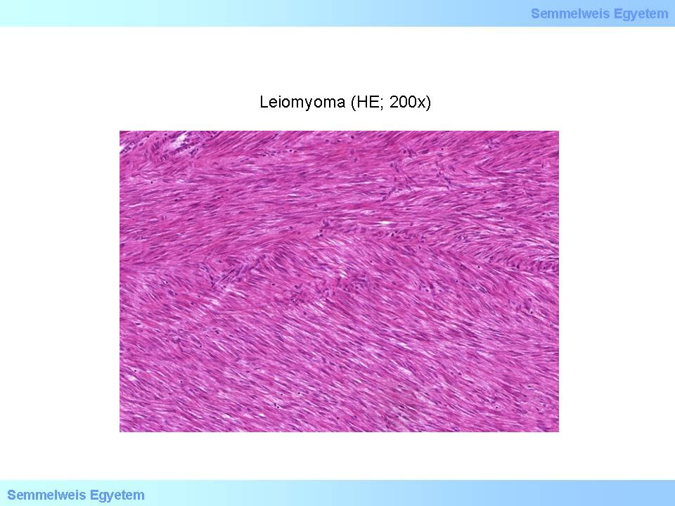
1st microphoto: Leiomyoma. The monotonous, spindle-shaped smooth muscle cells form characteristic spindle-shaped bundles remiscent of fish schools (HE; 200x) (From the photo archive of the Semmelweis University 2nd Institute of Pathology – collected by Ildikó Szirtes).
|
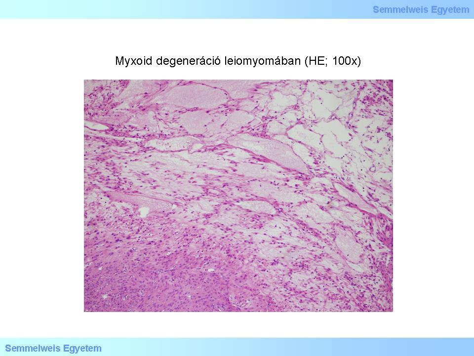
2nd microphoto: Myxoid degeneration in a leiomyoma. Pale, slightly basophile material accumulates in the stroma due to hypoxia. (HE; 100x) (From the photo archive of the Semmelweis University 2nd Institute of Pathology – collected by Ildikó Szirtes).
|

2nd macrophoto: Leiomyoma. In the single, massive leiomyoma deforming the uterine corpus a typical degenerative phenomenon: irregular area with myxoid density loss can be observed. (From the photo archive of the Semmelweis University 2nd Institute of Pathology – collected by Attila Kovács and Ildikó Szirtes).
|
If the tumour is totally calcified, ossified, the condition is called leiomyoma petrificatum. The tumour surface is filled with hard, crater-like recesses, which results in a meteorite-like look of the tumorous node. The so-called red degeneration is an unusual, but not rare form of leiomyomal necrosis, developing mainly during pregnancy (3rd micropicture). The condition can clinically be accompanied by pain and fever. In these cases the cut surface of the tumour is reddish, with a slightly softened substance. Histologically in some cases the leiomyoma is expressly stroma-poor and rich in cells (so-called cellular leiomyoma). In these cases the macroscopic picture also differs from the regular one: the cut surface is brown-yellowish, and the tumour tissue is also softer than usually.
|

Please take a look at the photo!
|
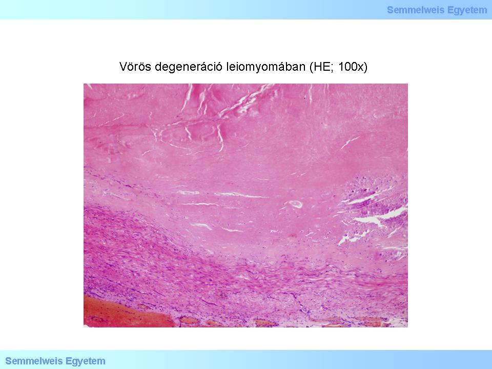
3rd microphoto: Red degeneration in leiomyoma. The smooth muscle node is almost totally replaced by dark, eosinophil, unifocal calcifying hyalinic necrosis
(HE; 100x) (From the photo archive of the Semmelweis University 2nd Institute of Pathology – collected by Ildikó Szirtes).
|
The cellular picture can vary between broad limits without the meaning of malignancy. Sometimes large, bizarre, hyperchromatic nuclei can occur in the so-called bizarre leiomyomata (4th microphoto), but if the mitotic rate is below 10 mitoses / 10 high power field (HPF) the tumour is not considered as a sarcoma yet. In summary, histological types of leiomyoma include:
-
1. cell-rich,
-
2. mitotically active,
-
3. leiomyoma with bizarre cells
-
4. epithelioid leiomyoma.
It is important to note that uterine leiomyomata are not considered as precursors of leiomyosarcoma. Their clinical symptoms depend on their location, size and can be very diverse: asymptomatic conditions, infertility, irregular cycle, pelvic space-occupation, etc.
|

Please take a look at the photo!
|
4th microphoto: Bizarre cell leiomyoma. In the cellular substance focally a few enlarged cells with an irregular form and a hyperchromatic nucleus can be observed as well, among the usually monomorph cells (HE; 200x) (From the photo archive of the Semmelweis University 2nd Institute of Pathology – collected by Ildikó Szirtes).
|
|