| |
I./1.1.: Anatomy of the kidney and ureter
I./1.1.1.: General description
|
 |
A paired organ, the kidney is situated retroperitoneally in the abdominal cavity. It has anterior and posterior surfaces, connected by a more convex lateral margin and an upright medial margin. Midway down the latter, there is the renal hilum. Around the hilum, the medial margin is separated into a thicker posterior lip and a thinner anterior lip. Both margins meet at the superior and inferior poles of the kidney.
|

Have a look at the image / figure!!
|

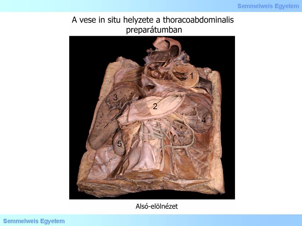
1. fotó: A bal vese in situ helyzete a thoracoabdominalis preparátumban – Pápai Zsolt, Molnár Attila és Balogh Attila: (1) Szív; (2) Gyomor; (3) Máj; (4) Lép; (5) Jobb oldali vese
|

2. fotó: A bal vese hilusa – Pápai Zsolt, Molnár Attila és Balogh Attila: (1) Arteria renalis; (2) Urether
|
I./1.1.2.: Fixation of the kidney and linked structures
|
 |
Typically, the human kidney has a smooth surface, however, in embryonic and fetal ages, it is segmented like a mulberry (ren lobatus). Such segmentation persists longer in certain species (e.g. bears, otters). Characteristic for visceral organs, the surface of the kidney is covered by a fibrous capsule, surrounded by adipose capsule and renal fascia. The latter is subdivided into prerenal fascia (of Gerota) and retrorenal fascia (of Zuckerkandl). The two sheets join each other only laterally, on the internal surface of the abdominal wall, and above, in front of the crura of diaphragm. Thus, the renal fascia forms a common envelope for both kidneys, also containing the suprarenal glands, abdominal aorta, inferior vena cava and adjoining renal and suprarenal blood vessels. This envelope is open below for passage of vessels and the ureter.
A robust pararenal fat pad (corpus adiposum retrorenale), as distinct from the adipose capsule is situated between the fascia of Zuckerkandl and the transversalis fascia of the abdomen. All three capsules play an important role in the fixation of kidney. Connective tissue ligaments are stretched between the inner fibrous capsule and the outer fascia, and the tension of these ligaments is maintained by the adipose capsule. At the same time, the renal fascia is adherent to the transversalis fascia behind and the parietal peritoneum in front. Both the fibrous and adipose capsules also extend through the hilum into the renal sinus. Thus the latter is filled with adipose tissue, into which the lesser and major calyces, renal pelvis (pyelon), interlobar branches of renal artery and vein, as well as lymph vessels and autonomic nerves are embedded.
I./1.1.3.: Urinary passages
|
 |
In the human kidney, 3-4 minor calyces join to form one major calyx, the total number being typically 2-3 major and 9-12 minor calyces. The major calyces (usually one upper and one lower) open into the renal pelvis, which is usually a dorsoventrally flattened sac but it may also adopt a rounded or narrow and tubular form.
The ureter arises from the renal pelvis and, following a retroperitoneal course, it ends in the urinary bladder (vesica urinaria). The following segments can be distinguished: abdominal and pelvic. Border beween the two segments is signified by the common or external iliac artery. Thickness of the ureter is not uniform, with three important constrictions. The first constriction is found at the pyeloureteric border, the second at the border beween the abdominal and pelvic segments, and the third at its entry to the bladder.
|

Have a look at the image / figure!!
|

3. fotó: A bal vese állománya – Molnár Attila és Balogh Attila: (1) Arteria renalis; (2) Urether; (3) Calix minor; (4) Cortex renalis; (5) Medulla renalis
|
I./1.1.4.: Vascular supply
|
 |
Vascular supply of the kidney comes from the paired renal arteries, emerging at the level of L1, often already divided into lobar branches prior to their entry to the hilum. Also occurring variations are accessory arteries, entering to the hilum but originating separately from the aorta, as well as so called polar arteries, which reach the superior or inferior pole directly. Of the latter, an inferior polar artery, usually crossing the ureter anteriorly, may also obliterate it.
Venous blood from both kidneys is returned to the inferior vena cava. The veins are situated slightly more caudally and ventrally to the arteries. Thus, the left renal vein crosses the abdominal aorta and, by contrast to the right renal vein, it also receives two stronger visceral branches: above the left suprarenal vein and below the ovarian or testicular vein.
Blood supply of the ureter originates from common sources together with neighbouring organs: renal, ovarian (testicular), common iliac and inferior vesical arteries.
|

Have a look at the image / figure!!
|

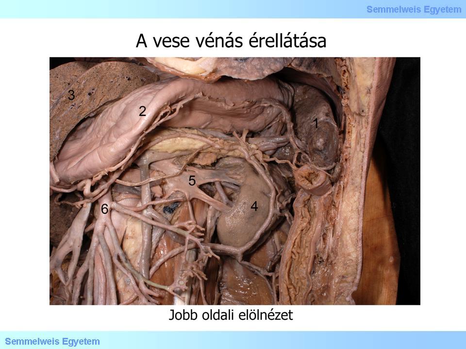
4. fotó: A bal vese vénás érellátása – Pápai Zsolt, Molnár Attila és Balogh Attila: (1) Lép; (2) Gyomor; (3) Máj; (4) Vese; (5) Vena renalis; (6) Arteria mesenterica superior
|
I./1.1.5.: Topography of the kidney and the ureter
|
 |
The right kidney lies between the vertebrae L1-L3, whereas the left kidney is situated half a segment further up, between the Th12/L1 transition and the body of vertebra L3. Typically, in females the kidneys are shifted by half a vertebra in a caudal direction, as compared with males. Ventral abdominal projections of the right or left kidney are transected by rib 12 or ribs 11 and 12, respectively. Having said that, the position of the inferior and superior poles of kidneys tend to vary substantially, between the vertebrae Th10 and L5. The medial border of kidneys extends as far as the costal processes of lumbar vertebrae, whereas the lateral border extends to a variable degree, as far as the lateral edge of quadratus lumborum muscle. The renal hilum lies in the level of L1.
Owing to a convergence of superior poles, the longitudinal axes of kidneys are not exactly parallel to each other. Thus, the distance between the superior or inferior poles (on average) is 7 or 11 cm, respectively. Given their position relative to the psoas major muscle, the surfaces of the kidney are not parallel either, forming an obtuse angle facing backward, whereby the hila are directed forward and in a medial direction.
|

Have a look at the image / figure!!
|

1. rajz: Az egészséges vizeletkiválasztó rendszer: (1)Vesék, (2) Vena cava inferior, (3) Aorta, (4) Arteria mesenterica superior, (5) Arteria ilaca communis, (5) Ureter, (6) Vesica urinaria, (7) Venae renales, (8)Elölnézet.
|
Since the kidneys are retroperitoneal organs, they are related anteriorly to the parietal peritoneum and posteriorly to the psoas major and quadratus lumborum muscles. Near their superior pole, the kidneys come in contact with the suprarenal gland, which is also enclosed by the adipose capsule. The right kidney is related - partly via the suprarenal gland - to the visceral surface of right hepatic lobe (hepatic facet of kidney), in front of the hilum to the descending (second) part of duodenum (duodenal facet of kidney), and, in its lower third, to the right colic flexure (colic facet of kidney). Important peritoneal ligaments of the kidney are the hepatorenal and duodenorenal ligaments. Anterior surface of the left kidney is topographically related, at the superior pole to the stomach (gastric facet of kidney), at the inferior pole to the left colic flexure (colic facet), at the hilum to the pancreas and, more laterally, to the spleen (splenic facet).
Having emerged from the hilum, the ureter turns immediately downward and nearly touches the inferior pole while heading for the pelvis in a caudal and slightly medial direction. In its course, the ureter crosses behind the ovarian (testicular) artery and vein. Further caudally, at the pelvic transition, it crosses in front of the common iliac artery (right side) or the external and internal iliac arteries (left side). The root of mesentery crosses above both ureters. At the transition between the abdominal and pelvic segments, the ureter raises a peritoneal fold named the fold of Douglas (plica Douglasi). In the pelvis, the ureter crosses behind the uterine artery (in the female), or the vas deferens (in the male) before entering the bladder from dorsal and inferior, also coming in contact (in the female), with the lateral fornix of vagina.
|
|